Early detection is key to curing the deadliest cancer.
Written by John Ferrari
Lung cancer is the deadliest form of cancer, but advances in the detection, diagnosis and treatment of the disease are pushing back on its toll. Torrance Memorial is the South Bay’s leader in bringing those advances to patients.
According to the American Lung Association, there’s a new lung cancer diagnosis in the United States every two minutes, and every day the disease takes almost 360 lives. Despite these sobering statistics, there have been tremendous strides in the detection and treatment of lung cancer. While it remains the leading cause of cancer deaths, over the past five years for lung cancers diagnosed when they are less than 1 cm, the five-year survival rate is 92%. For cancers diagnosed when they are between 1-2 cm, the five-year survival rate is 83%.
The key is early detection, says John Abe, MD, a Torrance Memorial physician specializing in internal medicine, pulmonary disease and critical care medicine. “It’s all about the stage at which it’s caught and treated. The smaller the tumor, the more time we have to treat it and the easier it is to treat. Over the last 15 to 20 years there has been a significant change in that we are diagnosing more early-stage cancers. About 75% of lung cancers diagnosed with robotic bronchoscopy at Torrance Memorial are early Stage 1 or 2.”
“The earlier the stage, the better the outcome,” agrees Khalid Eltawil, MD, a Torrance Memorial physician specializing in internal medicine and pulmonary disease. “Stage 1 cancers can essentially be cured.”
One of the most important advances in early detection is the recognition that screening saves lives. “Every improvement in the successful treatment of specific cancers has been based on an improved screening test,” notes Clark Fuller, MD, Torrance Memorial director of thoracic surgery and co-director of the thoracic oncology program. “For a long time, lung cancer detection relied on chest X-rays and incidental findings [that is, detections resulting from chest scans made for other reasons]. Eventually, large-scale trials showed that screening certain demographics for lung cancer—primarily individuals with a history of smoking—improved survival rates by over 20% for those screened, and CT scans became the screening modality of choice. CT scans are very sensitive, though, so the question becomes, ‘What do you do with all this data?’”
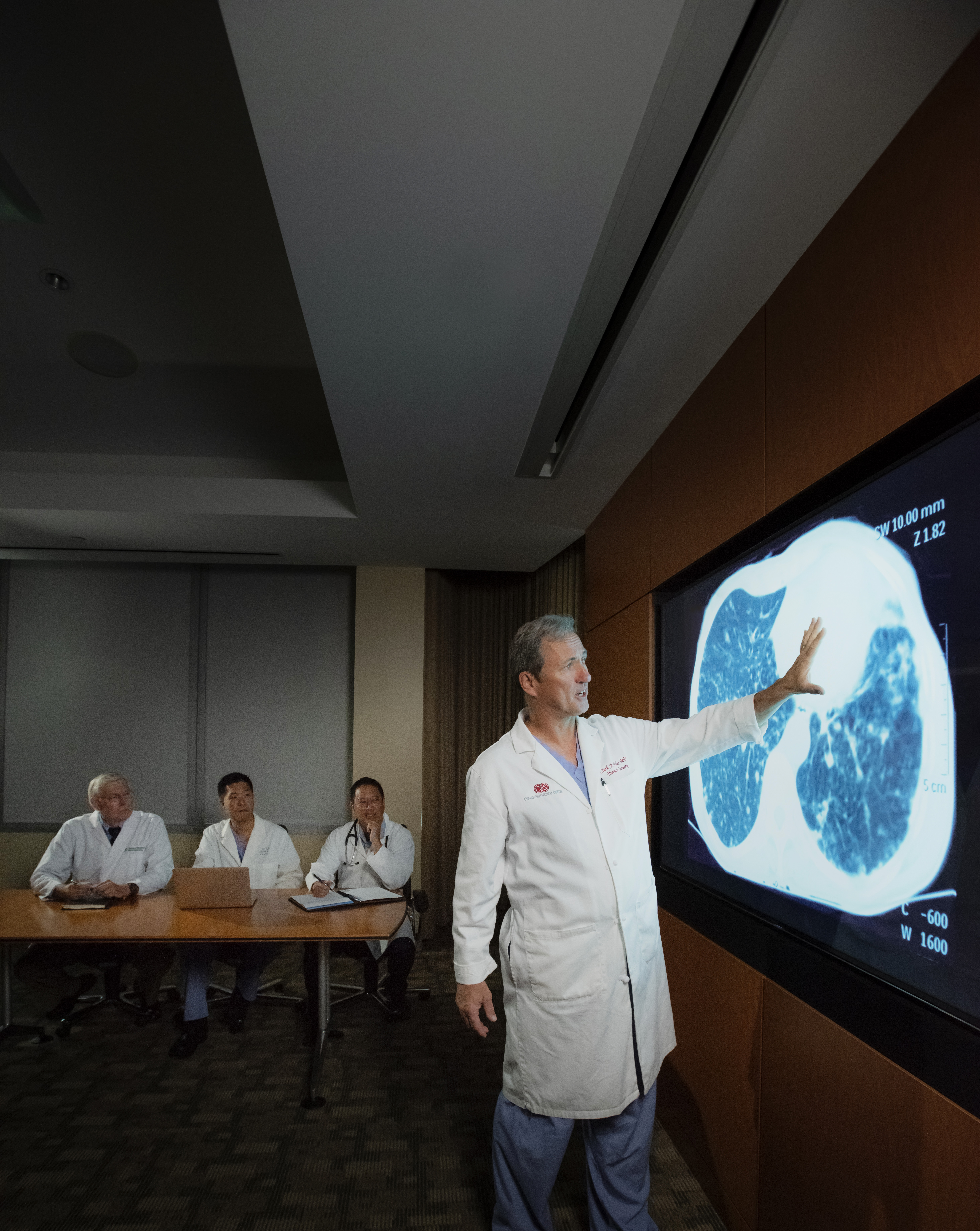
That’s where advances in medical imaging analysis come into focus. Torrance Memorial has adopted and integrated several imaging tools that use artificial intelligence learning—first to detect nodules on chest scans, and then to guide the diagnosis and treatment process. ClearRead, developed by Riverain Technologies, analyzes chest CT and X-ray scans to highlight nodules even smaller than 5 mm in size—well below the 2 cm threshold for the highest cure rates.
“When you look at a CT scan, the naked eye can sometimes miss tiny nodules,” Dr. Eltawil explains. “Riverain is able to remove the clutter and light up nodules so they’re easier to pick up.”
ClearRead integrates with another program, PowerScribe by Nuance Communications, to move from detection to diagnosis and treatment. “It puts together the likelihood of there being a nodule in a scan—not only to identify it more quickly but also to define a process on how to treat the nodule.”
The process then moves to Torrance Memorial’s lung nodule tumor board. “Every nodule is reviewed by the tumor board to determine the appropriate way to address it, whether that’s a biopsy, immediate removal, continued monitoring or some other treatment modality,” Dr. Eltawil says. “The process leads to improved workups of the findings, more timely diagnoses with higher confidence and the best treatment options.”
“This is where Ion comes in,” adds Dr. Fuller. The Ion bronchoscopy platform is a robotic-assisted system that allows Torrance Memorial pulmonologists to conduct minimally invasive biopsies of even the smallest suspicious nodules. The system uses CT imaging of a patient’s lung to create a 3D virtual model of the airways.
During the biopsy procedure this “road map” is presented along with real-time imagery of the patient, taken with a tiny video camera attached to a thin, maneuverable catheter, allowing the physician to precisely guide it to the nodule and retrieve a tissue sample. As the physician guides the catheter deeper into the lung tissue, the airway branches become smaller and smaller—going from the superhighway of the main airways, or bronchi, down to the one-lane roads of the smallest airways, the bronchioles.
There are roughly 25 descending sizes of these branchways. While a typical bronchoscopy procedure can reach into the four largest branchways, the Ion system allows physicians to reach into branchways more than 20 sizes smaller.
Dr. Abe puts the whole process in perspective: “With a CT scan, we can detect nodules that are very small, from 1 to 4 mm, and those are usually benign. The bigger they are, the more suspicious they are. Before we started using the Ion system, we could biopsy nodules as small as 15 to 20 mm. Now we can biopsy nodules 8 to 12 mm in size and confirm whether they’re benign or malignant.”
Once a nodule is definitively diagnosed as a malignant tumor, the board considers treatment options. “Chemotherapy is still an important treatment option, but if the tumor is caught early enough, it’s often not needed,” Dr. Abe says. “Radiation therapy is also a viable option, particularly for patients with conditions that make surgery risky. But for most patients, if we catch the cancer at an early stage, we can remove it and that’s the cure. It’s all about the stage at which it’s caught and treated.”
“That’s the key,” agrees oncologist and hematologist Andrew Horodner, MD, co-director of the thoracic oncology program at Torrance Memorial and chair of the hospital’s lung cancer tumor board. “There’s been a huge improvement in catching tumors early.”
Using early detection, diagnosis and minimally invasive surgical techniques, “we can get to small tumors we could never get to before,” he explains. “If we can remove the tumor, the patient is cancer-free. And for patients who do not want to have surgery or cannot have surgery for medical reasons, targeted radiation therapy can also produce excellent outcomes that are nearly equivalent to surgery. Our radiation oncology team has the most advanced technology available.”
Dr. Horodner adds that treatment options have expanded for patients diagnosed with non-small cell Stage 2 lung cancer—cases in which the tumor is larger than 4 cm and there may also be cancerous tissue in the thoracic lymph nodes. In approximately
1 in 4 such patients, systemic treatment—in this case, combined chemotherapy and immunotherapy—administered before surgery eliminates the cancer.
“We perform the surgery to be sure it’s completely gone, but this addition of preoperative treatment is a real paradigm shift. These patients have excellent long-term outcomes. We’re also using medication after surgery to improve treatment success. We’re always looking at how to improve those numbers. That’s the goal of our research effort with Cedars-Sinai: conducting studies to improve outcomes. We’re getting people the best, most comprehensive treatment.”
Torrance Memorial surgeons and physicians are continuing to improve lung cancer survival rates using these new tools for early detection and treatment. Dr. Fuller predicts the next leap will come from more early screenings.
“I think the criteria used for screening will change, especially among women,” he says. “Among women, there are more deaths from lung cancer than from breast, colon and uterine cancer combined—mostly because lung cancer cases tend to present at a later stage, especially among non-smokers. Oncologists and radiologists will review and adapt screening tools in response to this new epidemiological data. We’re also beginning to see that family history is important too, so there is a genetic component to lung cancer—that’s of vital importance. The more we can screen at-risk populations, the sooner we can catch lung cancer at Stage 1 and the more likely it is that we can cure that cancer. That’s a win-win.”
Spiral CT for high-risk patients: a new tool to catch lung cancer early
Early screening for lung cancer isn’t for everyone. “Roughly 85% to 90% of lung cancer cases are smoking-related,” notes John Abe, MD. “Overall, cases of lung cancer are trending down, but the number of new cases is still pretty high.”
That’s why early screening focuses on individuals with a history of smoking or past cancer diagnoses. More than 10 years ago, large-scale clinical trials confirmed the benefits of early screening.
"At Torrance Memorial, we were able to start an early screening program within a year of that published study,” says Dr. Abe. “We screen people who qualify as high-risk, and that’s primarily due to a history of smoking.”
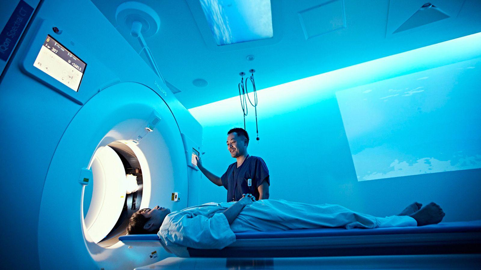
There’s a catch to early and repeated screenings, though. Lung cancer is detected using CT scans, and over time the radiation from CT scans can itself become a health risk. That’s why Torrance Memorial offers low-dose (also known as spiral or helical) CT scans for annual lung cancer screening of high-risk individuals.
CT scans are essentially 3D X-rays. Spiral CT scans rotate the X-ray tube around the patient in a spiral motion. The process is eight to 10 times faster than a traditional CT scan, and spiral CT scans are especially good at capturing small nodules. The X-ray dose from a spiral CT scan is about 1/3 less than the dose from a regular CT scan, and for high-risk individuals the benefits of the screening outweigh the risks, Dr. Abe says.
Studies have shown low-dose CT scans in high-risk patients improve the survival rate by 25%, adds Khalid Eltawil, MD. “The whole idea behind early screening is early detection because the earlier you detect lung cancer, the more survivable it is.”


